


Our
group
practice
offers
the
full
spectrum
of
diagnostics
and
therapy
for
cardiovascular
diseases
as
well
as
routine
check-ups.
We
have
five
modern
heart-and-vessel
ultrasound
units
at
our
disposal,
various
ECGs
(rest-,
exercise-stress
and
long-term
ECGs,
digital
X-
rays), and much more.
Our
new
MRI
equipment
has
been
in
operation
since
2004.
Magnetic
resonance
images
of
the
heart
have
many
advantages
over earlier standard exam methods, namely:
•
it
is
an
exam
that
has
no
effect
on
the
human
body.
There
are
no
radio
waves
emitted
(as
in
X-rays),
but
rather
magnetic
fields
-
thus
there
is
no
exposure to radiation.
•
the
images
provided
in
MR
are
in
very
sharp
focus,
enabling
the
physician
to
observe
the
heart
much
closer
and
more
accurately
than
is
possible
via
cardiac
ultrasound.
Moreover,
MR
enables
physicians
to
acquire
valuable
information
about
the
heart's
metabolism
(how
the
heart
muscle
uses
energy,
reflecting
its
"vitality").
The
doctors
can
tell
which
parts
of
the
heart
are
healthy
and
functioning
normally,
and
see
any
parts
containing
damaged
or
scarred
tissue
(i.e.,
after
a
heart
attack),
even
after
a
"silent
myocardial
infarction"
(a
heart
attack
the
patient
does
not
notice).
MR
also
provides
feedback
on
the
heart's
circulation
and
perfusion,
and
on
any
areas
suffering
from
ischaemia (oxygen deprivation).
Since
2007
we
have
benefited
from
a
further
new
diagnostic
tool,
namely
spiral
computer
tomography
(spiral
CT)
in
cooperation
with
Martha-Maria
Hospital.
This
method
allows
us
to
precisely
determine
the
degree
of
plaque
formation
in
the
coronary
arteries,
and
to
compare
those
findings
with
age-
and
gender-
specific
norms.
This
information
helps
us
make
a
reasonably
accurate
prognosis
of
a
patient’s risk of myocardial infarction.
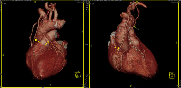
Under
the
right
conditions
(i.e.,
a
regular
heartbeat,
a
pulse
under
65/min,
atherosclerosis
that
is
not
too
advanced),
we
can
also
visualise
the
coronary
arteries
using
contrast
medium,
thereby
determining
which
if
any
vessels
are
stenotic
(partially
or
completely occluded or blocked by plaque).
For this exam, please contact our practice.
We
conduct
heart
catheter
examinations
(Prof.
Dr.
Jeserich,
Dr.
Kestler,
Dr.
Röther),
cardioversion
(Dr.
Kestler,
Dr.
Röther)
and
Pacemaker
implantations
(Dr.
Röther)
in
the
Dr. Steger Clinic, Nuremberg.
If
you
have
been
diagnosed
with
heart
and/or
vascular
disease,
or
should
you
wish
to
learn
more
about
your
risk
of
cardiovascular
disease,
our
team
of
specialists
and
consultants is at your service.
We
make
every
effort
to
ensure
that
diagnostic
reports,
findings,
and
therapy
recommendations
are
forwarded
to
the
referring
physician
(by
fax
within
24
hours,
by
mail
within
2
days)
as
quickly
as
possible
so
as
to
inform
the
patient
and
his
or
her
doctor
and
allow
ample
time
for
discussion.
We
kindly
remind
our
public
health
plan
patients
(Kassenpatienten)
to
bring
a
referral
note
(Überweisung)
along
when
ey
come
for
an
appointment.
Dr.
Röther
carries
out
vascular
exams
in
our
affiliated
practice
at
Schickenhof
6,
Sebalder
Hoefen.
Any
condition
concerning
varicose
veins,
thrombus,
swollen
legs
or
ankles,
or
circulatory
problems
in
the
legs,
arms
or
jugular
vein
can
be
handled.
We
can
also
carry
out
check-ups
and
follow-up
exams,
see
whether
you
suffer
from
plaque
formation
(and
if
so,
how
advanced
the
plaque
is),
and
whether
you
carry
an
increased
risk
of
stroke,
and many other issues.
Patients
with
lymph
oedema
(swelling)
are
treated
as
well
in
our
practice.
Special
lymph
drainage therapy can be performed.
Patients
with
lymph
oedema
(swelling)
are
treated
as
well
in
our
practice.
Special
lymph
drainage therapy is possible.
The
modern
technology
of
ergospirometry
allows
us
to
discover
what
is
causing
shortness
of
breath.
This
diagnostic
tool
is
also
useful
for
athletes
who
wish
to
determine
their
athletic
potential
(fitness
level),
or
for
check-ups.
We
also
offer
a
special
training
programme
whereby
the
body's
ability
to
burn
fat
is
analysed.
Arterial
stiffness
occurs
as
a
consquence
of
age
and
arteriosclerosis.
The
Puls
Wave
Analysis
(PWA)
is
done
during
the
blood
pressure
measurment
and
takes
about
5
minutes.
The
report
shows
the
age
of
your
blood vessels.
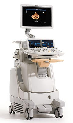
New
3D
cardiac
ultrasound
unit
with
various
diagnostic
capacities
including
colour-tissue
Doppler,
heart-volume
measurements,
3D-representation
of
the
heart in real time.
Click
on
the
picture
to
enlarge
the
image.
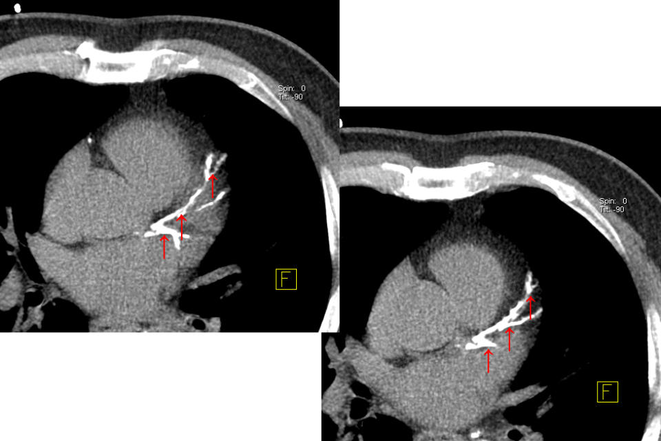
The early diagnosis of coronary plaque
(arrow) can potentially trigger a heart
attack (click to enlarge).
With this method, your coronary arteries
(arrow) are visualised non-invasively, that
is, without a catheter, thus revealing
stenosis (blockages) early (click to
enlarge).
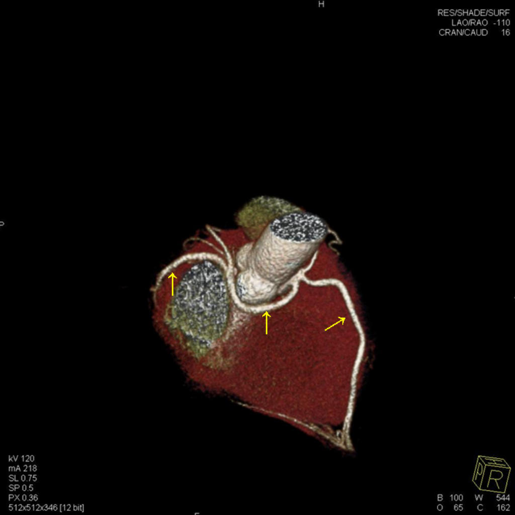
With this method, (arrow), bypasses can
be examined (click to enlarge).


Ergospirometric assessment
Since
2007
we
have
had
at
our
disposal
a
modern
cardiac
ultrasound
unit
with
three-
dimensional
imaging
capacity
in
certain
cases
(see image below).
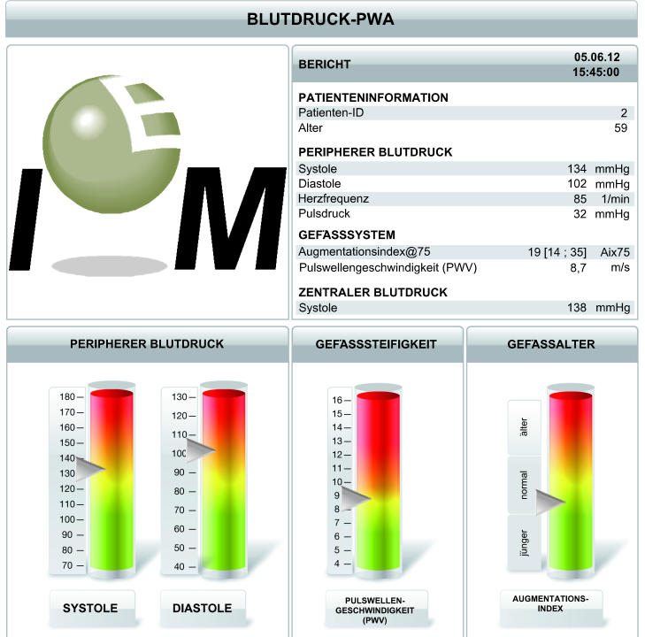
Puls Wave Analysis (PWA)

The web pages do not use cookies.